
Diagram of Human Lungs
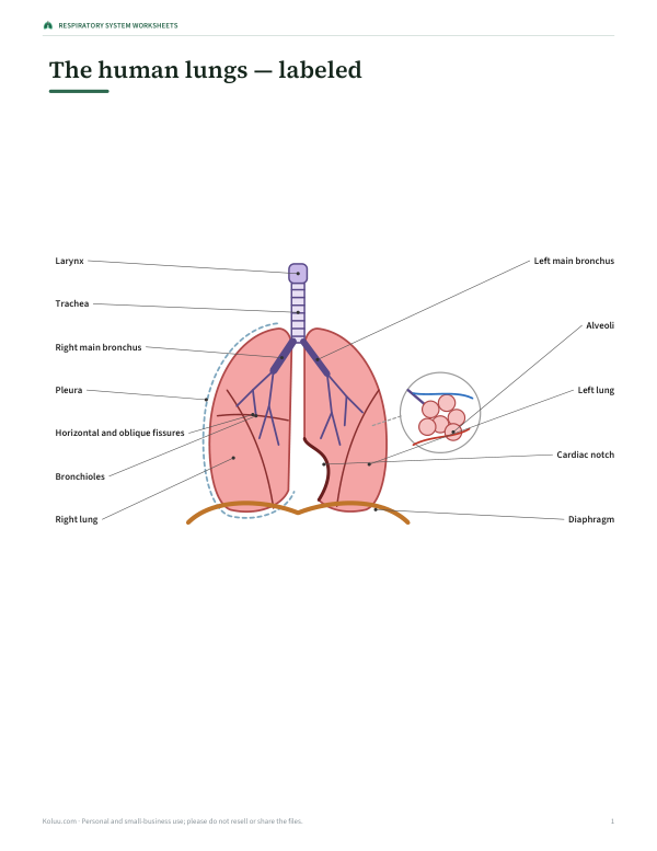
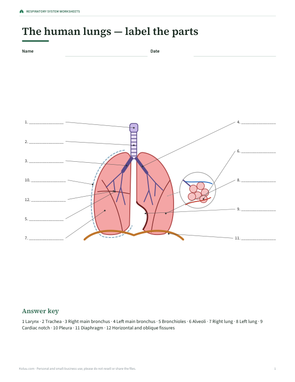
Your lungs bring oxygen into the body and remove carbon dioxide about 12 to 20 times a minute at rest. This interactive diagram shows the respiratory system from the front: the larynx and trachea, the branching bronchi, the lobes of each lung, the diaphragm and a close-up of the alveoli where gas exchange happens. Click any part to learn its job, press “Breathe” to see how the diaphragm moves, or test yourself.
- Free to use
- No sign-up
- Runs in your browser
Interactive lung diagram
Respiratory System Worksheets
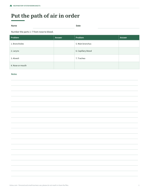
Printable respiratory system worksheets with answer keys: labeled and blank lung diagrams, a path-of-air sequencing activity, a gas exchange worksheet and a 12-question quiz.
- Labeled diagram (PDF)
- Blank diagram (PDF)
- Path of air (PDF, DOCX)
- 12-question quiz (PDF, DOCX)
Formats: PDF, DOCX. Instant download after payment (link valid 72 hours, up to 5 downloads). AI-assisted: the templates were drafted with AI help and reviewed and laid out before publishing.
$5.00 USD, one-time 4 files · about $1.25 each
- Instant download
- 7-day refund
- Secure checkout by Stripe
7-day money-back guarantee. Full refund within 7 days of purchase, even after you download.
Free sample: Lungs Blank (PDF, 65 KB)
Full refund within 7 days — see the refund policy and license.
Free printables
Single sheets you can print right now, no sign-up. The full pack adds every sheet in one download plus the editable files.
 Printable Respiratory System Quiz
Parts of the respiratory system
| Part | What it does |
|---|---|
| Larynx | The voice box at the top of the trachea. It contains the vocal cords and closes (with the epiglottis) when you swallow so food doesn’t enter the airway. |
| Trachea | The windpipe: a tube about 10–12 cm long held open by C-shaped rings of cartilage, carrying air from the larynx to the bronchi. |
| Right main bronchus | The branch of the trachea into the right lung. It is wider, shorter and more vertical than the left, so inhaled objects more often lodge here. |
| Left main bronchus | The branch of the trachea into the left lung; longer and narrower than the right because it passes under the arch of the aorta. |
| Bronchioles | Small airways branching from the bronchi, without cartilage, that carry air deeper into the lung and end in the alveoli. |
| Alveoli | Tiny air sacs — hundreds of millions of them — surrounded by capillaries. Oxygen diffuses into the blood and carbon dioxide diffuses out here. |
| Right lung | The larger lung, with three lobes (upper, middle and lower) separated by two fissures. |
| Left lung | The smaller lung, with two lobes (upper and lower) and a cardiac notch that makes room for the heart. |
| Cardiac notch | A hollow in the front edge of the left lung where the heart sits. |
| Pleura | Two thin membranes around each lung with a film of fluid between them that lets the lungs slide smoothly as you breathe. |
| Diaphragm | A dome-shaped sheet of muscle below the lungs. It contracts and flattens to draw air in, and relaxes to let air out. |
| Horizontal and oblique fissures | Grooves dividing the lungs into lobes: the right lung has both, the left lung only an oblique fissure. |
The path of a breath
- Air enters through the nose or mouth, where it is warmed, moistened and filtered.
- It passes the pharynx and the larynx, then down the trachea.
- The trachea divides into the right and left main bronchi, one for each lung.
- The bronchi branch again and again — about 20 to 23 times — into ever smaller bronchioles.
- The smallest airways end in clusters of alveoli, where oxygen passes into the blood and carbon dioxide passes out.
- Breathing out reverses the journey.
How breathing works
Breathing in is active: the diaphragm contracts and flattens, and the external intercostal muscles lift the ribs up and out. The chest cavity gets larger, pressure inside the lungs falls below the air pressure outside, and air flows in. Quiet breathing out is mostly passive: the muscles relax, the elastic lungs recoil, and air flows out. During exercise, extra muscles help with both. The “Breathe” button animates this movement in a simplified way.
Gas exchange in the alveoli
Each alveolus is a tiny sac with a wall only one cell thick, wrapped in a net of capillaries. Oxygen diffuses from the air in the alveolus into the blood, where it binds to haemoglobin in red blood cells; carbon dioxide diffuses the other way to be breathed out. Adult lungs contain several hundred million alveoli with a combined surface area often compared to a tennis court — roughly 50–100 square metres — which is what makes gas exchange so efficient.
Why the two lungs are different
The right lung has three lobes and the left lung two. The heart sits slightly to the left in the chest, so the left lung is smaller and has a cardiac notch in its front edge. The right main bronchus is wider and more vertical than the left, which is why an inhaled object such as a peanut is more likely to end up in the right lung.
How to use the interactive diagram
- Start in Labeled mode and click each part to read its function.
- Press “Breathe” to watch the lungs expand as the diaphragm moves down.
- Switch to Numbered mode to hide the names and test yourself or print a worksheet.
- Try “Spot the part” and the function quiz to check what you know.
Worked example: a classroom lesson
A science class starts with the labeled diagram to trace the path of air from the larynx to the alveoli. Students watch the breathing animation and compare it with a model lung made from a bottle, balloons and a rubber sheet. In the function quiz, many choose “Trachea” for the description of the bronchi, so the teacher highlights where the trachea divides. The lesson ends with the numbered diagram printed as an exit ticket.
Keeping lungs healthy
- Don’t smoke or vape, and avoid second-hand smoke — smoking is the leading cause of lung cancer and COPD.
- Stay active: exercise strengthens breathing muscles and improves fitness.
- Reduce exposure to air pollution, dust and fumes, and use protective masks at work where needed.
- Keep up with recommended vaccinations such as flu and pneumococcal vaccines.
- See a doctor about a cough lasting more than three weeks, breathlessness, coughing up blood or chest pain.
A note on the diagram
This is a simplified front view, not to scale. The heart, ribs and blood vessels are left out so the airways are easier to see, and the alveoli are shown as an enlarged close-up.
Frequently asked questions
What are the main parts of the lungs?
The bronchi, bronchioles, alveoli and the lobes, surrounded by the pleura, with the diaphragm below.
How many lobes does each lung have?
The right lung has three and the left lung two.
Where does gas exchange happen?
In the alveoli, tiny air sacs surrounded by capillaries.
What does the diaphragm do?
It contracts and moves down to draw air in, and relaxes to let air out.
Why is the left lung smaller?
It shares space with the heart and has a cardiac notch.
Can I print a blank lung diagram?
Yes, choose Numbered mode and print.